
 |
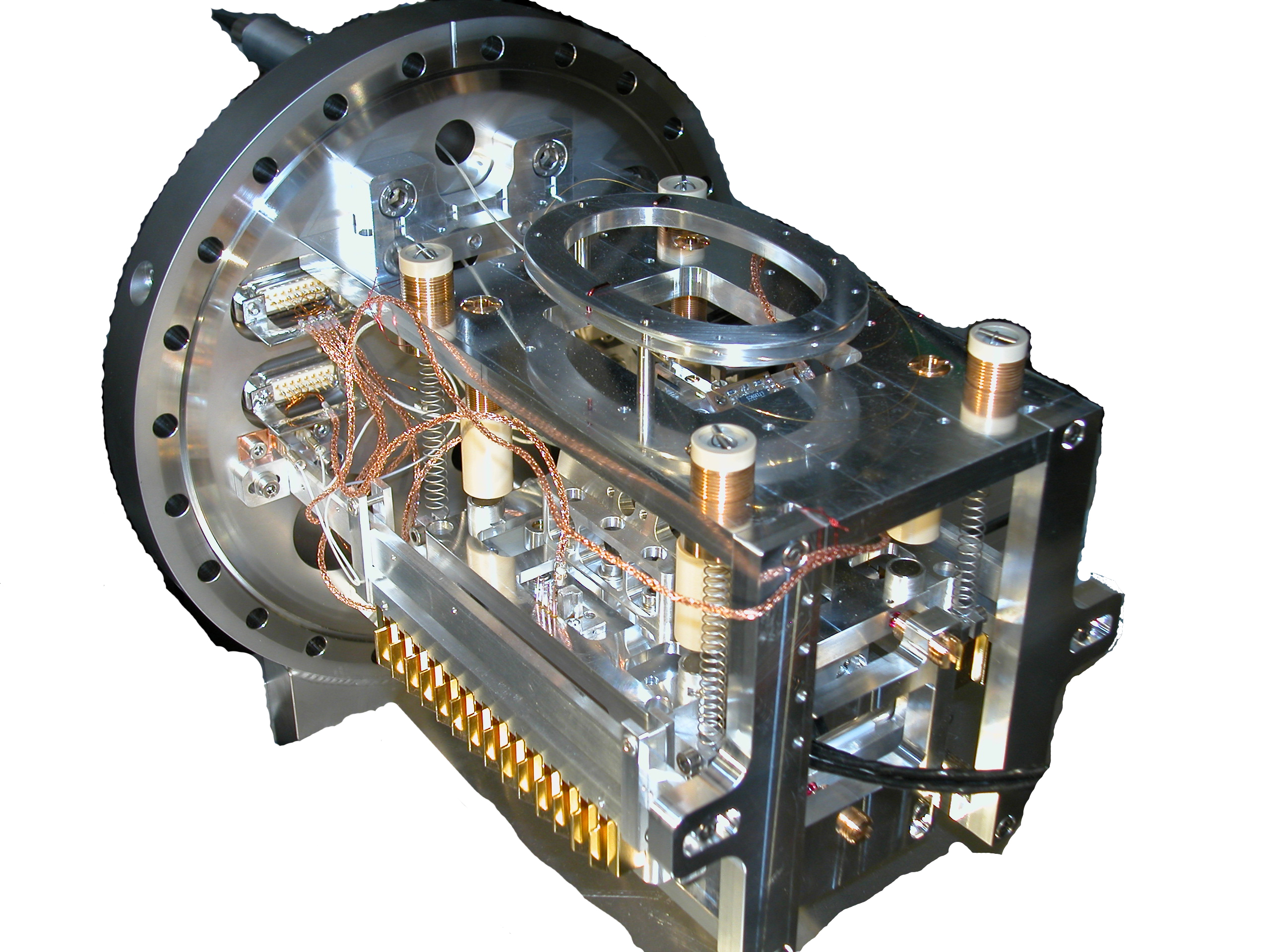
The resolution in Electrostatic Force Microscopy (EFM), a descendant of atomic force microscopy (AFM), has reached nanometer dimensions, necessary to investigate circuits in modern electronic devices. However, the characterization of conducting or semiconducting power devices with EFM methods requires an accurate and reliable technique from the nanometer up to the micrometer scale. For high force sensitivity it is indispensable to operate the microscope under high to ultra-high vacuum (UHV) conditions to suppress viscous damping of the sensor. Furthermore, UHV environment allows the analysis of clean surfaces under controlled environmental conditions. Because of these requirements we built a large area scanning probe microscope (SPM) operating in UHV conditions at room temperature allowing to perform various electrical measurements, like Kelvin Probe Force Microscopy (KPFM), Scanning Capacitance Force Microscopy (SCFM), Scanning Spreading Resistance Microscopy (SSRM), and also Electrostatic Force Microscopy (EFM) at higher harmonics. The instrument incorporates beside a beam deflection detection system a closed loop scanner with a scan range of 10µm in lateral 25µm in vertical direction as well as an additional light fibre feed-through for opto-electronic excitation measurements like surface photo voltage (SPV) detection. |
 |
||
| EFM on n-doped SiC calibrations layers. (a) schematic view, (b) KPFM with laser illumination (l=500±25nm), (c) 1st (d) 2nd and (e) 3rd harmonic of the periodic force. (f) a SSRM image. |
Publications related to this instrument:
| Kelvin probe force microscopy for material characterization Th. Glatzel, U. Gysin, and E. Meyer Microscopy, 71, (2022), i165-i173, pdf. |
| SPM Based Electrical Characterization Techniques for Semiconductors â Demonstrated for SiC Devices Th. Glatzel , U. Gysin, and E. Meyer International Symposium on Photonics and Electronics Science and Engineering 2016 (ISPESE 2016), 2016-03-11, Kyoto, (Japan). |
| Dopant imaging of power semiconductor device cross sections U.Gysin, E.Meyer, Th.Glatzel, G.Günzburger, H.R.Rossmann, T.A.Jung, S.Reshanov, A.Schöner, H.Bartolf Microelectronic Engineering, 160, (2016), 18-21, pdf. |
| Dopant imaging of Si and SiC structures using different SPM methods Urs Gysin; Ernst Meyer; Thilo Glatzel Swiss Nanoconvention 2015, 2015-06-30, Neuchâtel, (Switzerland), Abstract (PDF). |
| Two-Dimensional Carrier Profiling on Lightly Doped n-type 4H-SiC Epitaxially Grown Layers H. Rossmann, U. Gysin, A. Bubendorf, Th. Glatzel, S. Reshanov, A. Schöner, T. Jung, E. Meyer and H. Bartolf Material Science Forum, 821-823, (2015), 269-272, pdf. |
| Development of Power Semiconductors by Quantitative Nanoscale Dopant Imaging H. Bartolf, U. Gysin, H. Rossmann, A. Bubendorf, Th. Glatzel, T. Jung, E. Meyer, M. Zimmermann, S. Reshanov and A. Schoner IEEE 27th International Symposium on Power Semiconductor Dev, (2015), 281-284, pdf. |
| Large area scanning probe microscope in ultra-high vacuum demonstrated for electrostatic force measurements on high-voltage devices U. Gysin, Th. Glatzel, Th. Schmölzer, A. Schöner, S. Reshanov, H. Bartolf and E. Meyer Beilstein Journal of Nanotechnology, 6, (2015), 2485-2497, pdf. |
| Improving the Design of the Shield for the Electric Field in SiC-Based Schottky-Rectifiers and Ion-Implantation Cascades by SPM Dopant-Imaging H. Bartolf, U. Gysin, Th. Glatzel, H. Rossmann, T. Jung, S. Reshanov, A. Schöner and E. Meyer Microelectronic Engineering, 148, (2015), 1-4, pdf. |
| Design and performance of a combined secondary ion mass spectrometry-scanning probe microscopy instrument for high sensitivity and high-resolution elemental three-dimensional analysis T. Wirtz, Y. Fleming, M. Gerard, U. Gysin, Th. Glatzel, E. Meyer, U. Wegmann, U. Maier, A. H. Odriozola and D. Uehli Rev. Sci. Instrum., 83, (2012), 063702, pdf. |
| Combined SIMS-SPM Instrument For High Sensitivity And High Resolution Elemental 3D Analysis T. Wirtz, Y. Fleming, U. Gysin, Th. Glatzel, E. Meyer, U. Maier and U. Wegmann Microscopy and Microanalysis, 18, (2012), 888-889, pdf. |